| Table of Contents |  |
|
Case Report
| ||||||
| Successful surgical treatment for infectious giant bullae with mediastinal shift: A case report | ||||||
| Masaki Hashimoto1, Akifumi Nakamura1, Nobuyuki Kondo1, Seiki Hasegawa1 | ||||||
|
1Hyogo College of Medicine Department of Surgery, division of Thoracic Surgery.
| ||||||
| ||||||
|
[HTML Abstract]
[PDF Full Text]
[Print This Article]
[Similar article in Pumed] [Similar article in Google Scholar] |


| How to cite this article: |
| Hashimoto M, Nakamura A, Kondo N, Hasegawa S. Successful surgical treatment for infectious giant bullae with mediastinal shift: A case report. J Case Rep Images Surg 2016;2:80–83. |
|
Abstract
|
|
Giant bulla is defined as a large bulla that occupy over one-third of the thoracic cavity. A 40-year-old Japanese male presented with severe dyspnea and high fever. Radiological examination revealed that several fluid-filled giant bullae had expanded and occupied the majority of the right hemithorax. As a result, mediastinal shift to the contralateral side had occurred. Despite the administration of antibiotic therapy, his condition did not improve. An emergency operation involving resection of the walls of giant bullae and precise suturing of the base of these bullae was performed. After the operation, his condition showed immediate improvement, and the residual right lung was fully expanded. It is essential to make definitive closure of bronchial communication and preserve residual lung parenchyma in cases of rapidly expanding infectious giant bullae.
| |
|
Keywords:
Infectious bullae, Mediastinal shift, Surgical treatment, Thoracotomy
| |
|
Introduction
| ||||||
|
Giant bulla is defined as a large bulla that occupy over one-third of the thoracic cavity [1]. It is an uncommon syndrome that differs from common forms of bullous emphysema by affecting a younger population, most commonly young men. Most of affecting patients have a history of smoking cigarettes. Most of clinical symptoms such as hypoxia, dyspnea, and easy to fatigue are often seen, but some of patients are asymptomatic [2]. As enlarging giant bulla, occupying the entire hemithorax and collapsed residual lung parenchyma, which is so called vanishing lung syndrome, is a common occurrence. However, a complicated mediastinal shift is extremely rare. We present a case wherein mediastinal shift occurred due to infectious giant bullae. | ||||||
|
Case Report
| ||||||
|
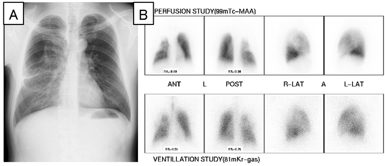
A 40-year old Japanese male was admitted as an emergency to our hospital due to severe dyspnea with high fever. He was a current smoker, with a smoking history of 30 pack-years and was under treatment for schizophrenia. He was referred to our hospital due to giant bullae on the right that was found four years previously. These giant bullae had continuously increased in size over time and occupied two-thirds of the right thoracic cavity one month before admission (Figure 1A). Although we recommended surgical resection of this giant bullae, the patient did not consent to the operation. At the emergency admission for severe dyspnea, a chest radiography revealed that the giant bullae on the right was filled with fluid (Figure 1B) and had expanded to occupy the majority of the right hemithorax. Moreover, pressure from it caused shifting of the mediastinum to the contralateral side. Chest computed tomography (CT) scan revealed that the residual right lung had collapsed. This was due to multiple giant fluid-filled bullae and aspiration pneumonia apparent in the left lung (Figure 2). The patient had a spike fever with severe hypoxia. Blood test results were as follows: C-reactive protein, 30.4 mg/dl, white blood cells 18680/µl and procalcitonin, 2.80 ng/ml. We diagnosed infected giant bullae with aspiration pneumonia, and antibiotic therapy (piperacillin/tazobactam) was immediately administered. However, his clinical symptoms and condition remained unchanged despite the antibiotic therapy. Therefore, emergency surgery was performed seven days after the admission. The operation was performed through a right thoracotomy in the right 5th intercostal space with a 12-cm incision. Initially, we incised the wall of the bullae and discharged the contents of pus and necrotic tissue. Thereafter, bullectomy was performed using an endoscopic stapling device. As we suspected communication between the bulla and a bronchiole, mattress imbricating sutures were placed at the base of the bullae. After the operation, hypoxia and high fever resolved immediately. On postoperative day 2 (POD-2), a chest drainage tube was removed and oxygen therapy was ended. A culture examination revealed Burkholderia cepacia. The antibiotic therapy was changed to doripenem, which was continued till POD-11. His postoperative course was uneventful, and he was discharged on foot on POD-25. Expansion of the residual right lung improved, and full expansion was achieved two years after surgery (Figure 3A). Lung scintigraphy showed that the right-to-left ratio was 46:54 (Figure 3B) . The patient was in good health at the last follow-up two years after surgery. | ||||||
|
| ||||||
| ||||||
| ||||||
|
Discussion
| ||||||
|
Mediastinal shift commonly presents with tension pneumothorax or massive pleural effusion. Although enlarged giant bullae could compress residual lung parenchyma, a complicated mediastinal shift is rarely observed. In this case, intrabullous effusion was produced by an active infection, and this induced the giant bullae to rapidly expand. Consequently, severe mediastinal shift occurred. Indication of surgical intervention for giant bulla is considered as patient's condition. Surgical intervention is usually performed in patients with dyspnea or complications such as pneumothorax, prolonged air leakage after tube insertion [2] [3], and is also rarely performed in patients with bleeding, infection, or cancer associated giant bullae [4]. The goal of surgical intervention for patient with giant bullae is whom medical treatment has failed and to resolve complications while preserving lung function [4]. Administration of systemic antibiotics is the most common therapeutic approach against infectious bullae [5]. However, in cases of infectious giant bullae, medical treatment is often unsuccessful because of the poor communication between the bullae and the bronchial tree [6]. Complicated infection within the giant bullae was one of the indications for surgical intervention in this case. The surgical intervention technique of choice for infectious bullae is usually bullectomy, usually performed via video-assisted thoracoscopic surgery (VATS) [7] [8]. As most patients with infectious bullae are in poor condition, VATS bullectomy is more suitable as it is less invasive than conventional open thoracotomy. However, in this case, we did not select VATS bullectomy because bronchial communication was suspected by the complicated aspiration pneumonia, and the border between normal lung parenchyma and the bullous wall was not clearly defined. We performed the bullectomy with definitive closure of bronchial communication and preserved the residual lung parenchyma via open thoracotomy. As a result of this operation, the patient's right lung was fully expanded, and his pulmonary function recovered to a normal range. Video-assisted thoracoscopic surgery bullectomy is less invasive and is an effective surgical approach for infectious giant bullae. However, it is not suitable in cases such as this one. In this case, it was necessary to remove the bullous wall and make definitive closure of bronchial communication, even if this required an open thoracotomy. | ||||||
|
Conclusion
| ||||||
|
We concluded that it is essential to make definitive closure of bronchial communication and preserve residual lung parenchyma in cases of rapidly expanding infectious giant bullae. | ||||||
|
References
| ||||||
| ||||||


|
[HTML Abstract]
[PDF Full Text]
|
|
Author Contributions
Masaki Hashimoto – Substantial contribution to conception and design, Acquisition of data, Drafting the article, Revising it critically for important intellectual content, Final approval of the version to be published Akifumi Nakamura – Substantial contribution to conception and design, Acquisition of data, Drafting the article, Final approval of the version to be published Nobuyuki Kondo – Substantial contribution to conception and design, Acquisition of data, Drafting the article, Revising it critically for important intellectual content, Final approval of the version to be published Seiki Hasegawa – Substantial contribution to conception and design, Drafting the article, Revising it critically for important intellectual content, Final approval of the version to be published |
|
Guarantor of submission
The corresponding author is the guarantor of submission. |
|
Source of support
None |
|
Conflict of interest
Authors declare no conflict of interest. |
|
Copyright
© 2016 Masaki Hashimoto et al. This article is distributed under the terms of Creative Commons Attribution License which permits unrestricted use, distribution and reproduction in any medium provided the original author(s) and original publisher are properly credited. Please see the copyright policy on the journal website for more information. |
|
|