|
|
Case Report
| ||||||
| Robotic repair of Amyand's hernia: A case report | ||||||
| Thomas J. Shaknovsky1, Frederick Sabido2, Dooniya Shaikh3, Phillip L. Rosen4 | ||||||
|
1DO, Southeast Alabama Medical Center/Alabama College of Osteopathic Medicine, General Surgeon/Clinical Professor, Dothan, AL USA.
2MD FACS, Richmond University Medical Center, Director of Minimally Invasive Surgery Fellowship, Staten Island, NY USA. 3BS, Richmond University Medical Center, Medical Student, Staten Island, NY USA. 4MD, Richmond University Medical Center – General Surgery Resident, Staten Island, NY USA. | ||||||
| ||||||
|
[HTML Abstract]
[PDF Full Text]
[Print This Article]
[Similar article in Pumed] [Similar article in Google Scholar] |


| How to cite this article: |
| Shaknovsky TJ, Sabido F, Shaikh D, Rosen PL. Robotic repair of Amyand's hernia: A case report. J Case Rep Images Surg 2016;2:92–96. |
|
Abstract
|
|
Introduction:
Inguinal hernia repair is the most common procedures in general surgery. Contents within the hernia sac are variable and in rare cases, the hernia sac contains the vermiform appendix termed Amyand's Hernia. While laparoscopic technique is the most accepted approach, few prospective studies have been completed comparing laparoscopic versus robotic techniques for hernia repair and none in the setting of an Amyand's hernia. We describe the first documented robotic approach to the repair of Amyand's hernia.
Case Report: A 40-year-old male presented to outpatient surgery clinic with complaint of right scrotal mass. He was diagnosed with right inguinal hernia and was electively scheduled for robotic repair with mesh. Intraoperatively, type 1 Amyand's hernia was discovered. Using robotic technique the hernia was reduced and successfully repaired with mesh. Patient postoperative course was unremarkable. Conclusion: Amyand's hernia is a rare subtype of inguinal hernia containing vermiform appendix within the hernia sac. Management of Amyand's hernia is surgery and extent of intervention is dictated by pathology of the vermiform appendix. While open and laparoscopic techniques have been well documented, robotic approach is safe, effective and provides a surgeon with a unique advantage in repair of Amyand's hernia. The robotic platform Da Vinci Surgical System's 3D high definition imaging coupled with advanced degree of dissection control provides a surgeon with a unique advantage over open and laparoscopic techniques. Robotic approach to repair of Amyand's hernia is safe and effective. | |
|
Keywords:
Amyand's hernia, Appendectomy, Incarcerated Amyand's hernia, Robotic herniorrhaphy, Robotic inguinal hernia
| |
|
Introduction
| ||||||
|
Inguinal hernia repair is the most common procedures in general surgery. However, the contents within the hernia sac are variable. In rare cases, the hernia sac contains the vermiform appendix. This is termed Amyand's Hernia after Claudius Amyand, an English surgeon who performed the first appendectomy on an eleven-year-old boy with a perforated appendix inside an inguinal hernia sac in 1735 [1] [2]. Usually, these are discovered as an incidental finding intraoperatively. The incidence of a normal appendix within the inguinal hernia sac is estimated to be 1% and that of an inflamed appendix to be 0.1% [2]. Treatment includes herniorrhaphy with or without appendectomy and/or mesh repair depending on the vermiform appendix's inflammation status, the patient's general condition, and other factors [3]. There are current classifications to guide physicians proposed by Losanoff and Basson in 2007 [4] [5] (Table 1). There are, however, few guidelines on the approach to these situations with laparoscopic or robotic methods. In a case series done by Sahu et al., three patients with Amyand's hernia underwent laparoscopic transabdominal preperitoneal hernioplasty without appendectomy. Investigators found that laparoscopic repair, in the setting of Amyand's hernia, has a number of advantages including better visualization of the sac and the ability to inspect and repair a left sided inguinal hernia if present [6]. In 2000, robotic surgery platform da Vinci was the first robotic surgery platform approved by Food and Drug Administration (FDA) for general laparoscopic surgery. In the years to follow, this novel technology quickly penetrated all aspects of surgical fields including general surgery. The robotic platform da Vinci Surgical System's 3D high definition imaging coupled with advanced degree of dissection control provides a surgeon with a unique advantage over open and laparoscopic techniques. This technology although not specific to the setting of hernias, has already proven to improve precision and dexterity, allowing surgeons to perform various operations that were traditionally not amenable to minimal access techniques [7]. A few prospective studies have been completed comparing laparoscopic versus robotic techniques for hernia repair and none in the setting of an Amyand's hernia. Thus as a start, this case report will document the first known documented robotic approach to repair a type 1 Amyand's hernia. | ||||||
| ||||||
|
| ||||||
|
Case Report
| ||||||
|
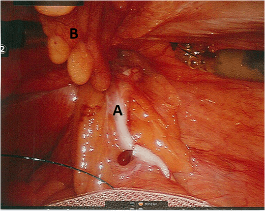
A 40-year-old male presented to the outpatient surgery clinic with a right sided scrotal mass which was present since childhood. Patient admitted that it progressively become enlarged but never became firm, irreducible, erythematous, or exquisitely painful. He denied changes in bowel habits, nausea, vomiting, fever, or chills. He did, however, complain that he could feel his bladder "slip into the swelling" on occasion which caused him difficulty in urination. This was not associated with any dysuria, pyuria, or hematuria. The patient had a past medical history significant for obstructive sleep apnea on a CPAP machine at home, obesity, and borderline hypertension. His only surgical history was a childhood tonsillectomy. He was not on any medications and has allergies to penicillin. He denied smoking or drinking but admitted to occasional marijuana use approximately once every two weeks. On physical examination, he presented with a reducible right inguinal hernia extending into his scrotum. There was no erythema or tenderness. The testicles were present in the scrotal sacs bilaterally. The patient was scheduled for an elective robotic assisted laparoscopic right inguinal hernia repair with mesh, possible bilaterally, in the ambulatory setting. Preoperative labs were within normal limits. Standard antiseptic technique was used to prep the patient and a total of three port site incisions were made for the da Vinci robot arms to dock and enter. The patient was placed in trendelenburg position and a robotic 8-mm trocars were placed in each anterior axillary line and a 30-degree camera was placed in the up position in the umbilical port. The robot was docked in the standard fashion. The right inguinal region was visualized and incarcerated inguinal hernia was noted. Once hernia contents reduced, a non-inflamed appendix and portion of the cecum was seen in the hernia sac. Type 1 Losanoff and Basson classification Amyand hernia was noted (Figure 1) and (Figure 2). The appendix, cecum, and pericolonic fat were reduced into the abdomen and the peritoneum was scored from the medial umbilical fold laterally to the anterior superior iliac spine. A preperitoneal flap was created by dissecting the peritoneum away from the spermatic cord vas deferens anteriorly with the hook electrocautery. A 3D max mesh and a 6-inch V-Loc suture were introduced intraperitoneally under direct visualization. The mesh was used to cover the preperitoneal space overlying the direct and indirect inguinal spaces. No direct hernia was observed. The indirect space and rest of the potential hernia sites were covered with mesh with adequate coverage. The mesh was tucked into the small peritoneal pocket and appeared secure without evidence of migration. The peritoneal flap was closed with a running 6-inch V-Loc suture. Patient was extubated in the OR and discharged home the same day after a non-complicated postoperative course. | ||||||
| ||||||
| ||||||
|
Discussion
| ||||||
|
Robotic surgery has progressed within the past 30 years from the Puma 560 used in 1985 by Kwoh et al. to perform neurosurgical biopsies to the NASA influenced da Vinci surgical system used today [8] [9]. This system is a complex of four arms controlled by a remote console usually in the same room as the patient. Through small incisions, the da Vinci robot features a magnified 3D high-definition visualization of the operative field and allows precise wristed movements with tiny instruments that can bend and rotate far better than the human hand. From their inception, surgical robots have extended the capabilities of human surgeons beyond the limitations of laparoscopic surgery. Currently there are only some small scale studies comparing the two techniques, such as that done at Baptist Health South Florida by Dominguez et al. This single institution study was a retrospective chart review of a total of 123 robotically repaired hernias, 45 of which were bilateral hernias. Researchers concluded that robotic transabdominal preperitoneal (TAPP) inguinal hernia repair is evidenced to be a safe and adaptable approach that allows surgeons to perform this procedure in more complex cases such as those involving incarcerated and/or recurrent hernias [10]. Furthermore, early studies have shown that da Vinci ventral hernia surgery has a lower rate of recurrent hernia as well as a lower rate of pain, and lower rate of switching to open [11] [12] [13]. In our case of Amyand's hernia, the precision and enhanced visualization provided by the da Vinci system allowed easy manipulation of the vermiform appendix and placement of mesh without trauma to the appendix. This is especially important considering the fact that any trauma endured by the appendix while it is being reduced can increase the risk of appendicitis developing postoperatively [14]. Local trauma while handling the appendix could potential transform classification of Amyand's hernia from type 1 to type 4, thus complicating treatment and intervention [15] [16]. | ||||||
|
Conclusion
| ||||||
|
Amyand's hernia, the presence of the appendix within the hernia sac, is a rare subtype of inguinal hernia and typically is an incidental finding. While there is no consensus on the ideal method for repair, Losanoff and Basson classification provides guidance on management. Robotic surgery is a relatively new field of the minimally invasive surgery spectrum. The superior anatomical imaging of the 3D high definition robotic endoscope coupled with the advanced dexterity, surgical control and degree of motion affords the surgeon a unique advantage over open and laparoscopic surgery. Our experience is the first documented case of robotic repair of Amyand's hernia and demonstrates that this is a safe and viable approach. | ||||||
|
References
| ||||||
| ||||||
|
Suggested Reading
| ||||||


|
[HTML Abstract]
[PDF Full Text]
|
|
Author Contributions
Thomas J. Shaknovsky – Substantial contributions to conception and design, Acquisition of data, Analysis and interpretation of data, Drafting the article, Revising it critically for important intellectual content, Final approval of the version to be published Frederick Sabido – Analysis and interpretation of data, Revising it critically for important intellectual content, Final approval of the version to be published Dooniya Shaikh – Analysis and interpretation of data, Revising it critically for important intellectual content, Final approval of the version to be published Phillip L. Rosen – Analysis and interpretation of data, Revising it critically for important intellectual content, Final approval of the version to be published |
|
Guarantor of submission
The corresponding author is the guarantor of submission. |
|
Source of support
None |
|
Conflict of interest
Authors declare no conflict of interest. |
|
Copyright
© 2016 Thomas J. Shaknovsky et al. This article is distributed under the terms of Creative Commons Attribution License which permits unrestricted use, distribution and reproduction in any medium provided the original author(s) and original publisher are properly credited. Please see the copyright policy on the journal website for more information. |
|
|
|
About The Authors
| |||
| |||
| |||
| |||
| |||