 |
|
Case Report
| ||||||
| Barium-induced allergic appendicitis: A case report | ||||||
| David L. Manuel1,4, Miriam Neufeld2,5, Laura M. Piechura2,7, Qin Huang3,6, Gentian Kristo2,7 | ||||||
|
1Department of Radiology, Veterans Affairs Boston Healthcare System, Boston, MA, USA
2Department of Surgery, Veterans Affairs Boston Healthcare System, Boston, MA, USA 3Department of Pathology, Veterans Affairs Boston Healthcare System, Boston, MA, USA 4Department of Radiology, Boston Medical Center/Boston University, Boston, MA, USA 5Department of Surgery, Boston Medical Center/Boston University, Boston, MA, USA 6Department of Pathology, Boston Medical Center/Boston University, Boston, MA, USA 7Department of Surgery, Brigham and Women’s Hospital/Harvard Medical School, Boston, MA, USA | ||||||
| ||||||
|
[HTML Abstract]
[PDF Full Text]
[Print This Article]
[Similar article in Pumed] [Similar article in Google Scholar] |


| How to cite this article: |
| Manuel DL, Neufeld M, Piechura LM, Huang Q, Kristo G. Barium-induced allergic appendicitis: A case report. J Case Rep Images Surg 2017;2:25–28. |
|
ABSTRACT
|
|
Appendicitis after barium administration occurs rarely and its underlying pathology remains undetermined. We present a case where acute appendicitis presented as an allergic reaction to barium sulfate. A 55-year-old male presented with nausea and severe right lower abdomen and right groin pain seven days after a barium swallow study. Computer tomography revealed retained barium in the appendix without any signs of inflammation. The patient underwent a laparoscopic appendectomy with resolution of his symptoms. Histopathologic examination demonstrated eosinophilic infiltration of the muscularis propria, consistent with acute eosinophilic appendicitis as a result of type I hypersensitivity reaction to barium. The recognition of potential risk of barium-induced allergic appendicitis is important for timely diagnosis. |
| Keywords: Allergic appendicitis, Barium Sulfate, Colorectal Surgery, Eosinophilic appendicitis |
|
INTRODUCTION
| ||||||
|
Barium-induced appendicitis is a rare complication after enteric barium examinations and was first reported as a case in 1954 [1]. The time span between barium study and onset of appendicitis ranges from a few hours to a few years [2] with the highest risk being within two months after barium administration [3]. Although the retained barium in the appendix is generally thought to form a barium-coated fecalith (barolith) leading to luminal obstruction and appendicitis [4], the pathophysiology of barium-induced appendicitis remains unclear. We present our experience with this rare surgical scenario in this report. | ||||||
|
CASE REPORT
| ||||||
|
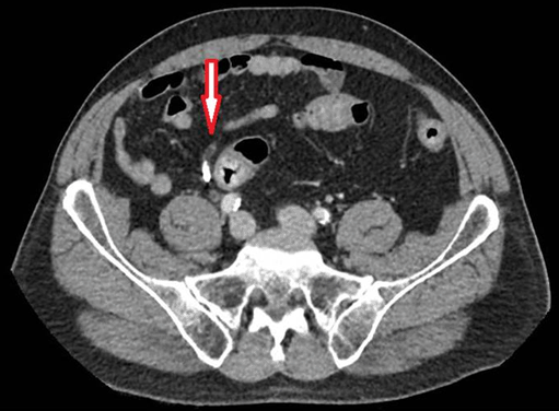
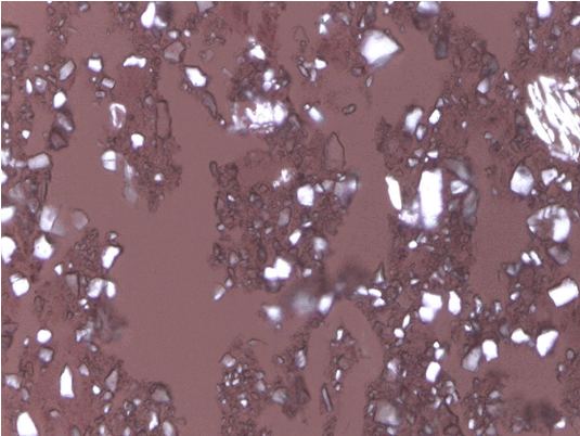
A 55-year-old male with dysphagia underwent an outpatient barium swallow study at an outside institution. A few hours later he developed nausea and severe pain in the right lower quadrant of the abdomen, prompting him to present at the same outside institution’s emergency department for an evaluation. He was admitted for overnight observation after a computed tomography scan of the abdomen and pelvis which was unable to provide a diagnosis. Six days later, the patient presented to our emergency department with nausea and severe right lower quadrant abdominal pain radiating into the right groin. Physical examination revealed significant tenderness in the right lower quadrant of the abdomen and over the right groin, without any evidence of groin hernias. The patient was afebrile, with normal leukocyte counts. A computed tomography scan of the abdomen and pelvis was obtained and showed retained barium in the appendix without any signs of appendicitis (Figure 1). After the patient was evaluated by consultant surgeons, he was taken immediately to the operating room for a laparoscopic exploration of the abdomen with the presumptive diagnosis of barium-induced appendicitis. At surgery, only minimal serosal hyperemia of the body of the appendix was found and an appendectomy was performed. Postoperatively, patient reported immediate and complete relief of his right lower quadrant and right groin pain. He was discharged to home 12 hours after the surgery and had an unremarkable recovery. Pathologic examination of the appendix showed a dilated appendiceal lumen without fecaliths, barium crystals in the mucosa of the appendix | ||||||
| ||||||
| ||||||
|
DISCUSSION
| ||||||
|
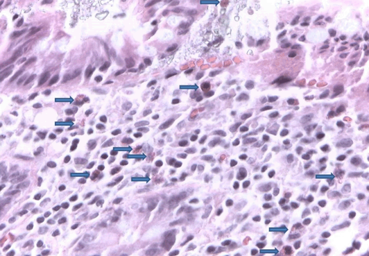
Herein, we report the case of a patient with barium-induced appendicitis, with symptoms starting hours after barium administration and correct diagnosis and treatment delayed for seven days. This indicates the importance of maintaining a high index of suspicion for barium-induced appendicitis in patients who present with symptoms of appendicitis after recent barium imaging.. Eosinophils are normally present in the lamina propria and submucosa, but not in the muscularis propria of the appendix [5]. The eosinophilic infiltration of the muscularis propria in our patient suggests a type I hypersensitivity reaction to barium or to any of the additives contained in the barium sulfate solution, including deflocculation agents, suspending agents, and flavoring agents [6]. If the mucosal injury caused by the eosinophils becomes infected by bacteria it leads to acute suppurative appendicitis, whereas in the absence of infection acute eosinophilic appendicitis (AEA) occurs [5]. Acute eosinophilic appendicitis is a known rare variant of appendix inflammation. The histologic hallmark of this entity is eosinophilic infiltration of the muscularis propria without neutrophilic infiltration [5][7], as was the finding in our patient. A count of > 10 eosinophils per high power field at microscopic examination is found in AEA [8]. In our case, there were about 150 eosinophils per high power field. Acute eosinophilic appendicitis has been related to multiple parasites including Strongyloides stercoralis [9], Schistosoma japonicum [10], and Entamoeba histolytica [11]. This case report is very significant because, to the best of our knowledge, it represents the first reported evidence of an acute eosinophilic appendicitis caused by retention of the barium sulfate solution in the appendix. | ||||||
|
CONCLUSION
| ||||||
|
Barium-induced appendicitis is a very rare clinical entity, but given the pervasive use of barium for enteric radiographic studies clinicians should recognize this potential risk to avoid delayed diagnosis and treatment. Histopathology is the gold standard for diagnosis of this rare condition. | ||||||
|
REFERENCES
| ||||||
| ||||||

|
[HTML Abstract]
[PDF Full Text]
|
|
Author Contributions
David L. Manuel – Substantial contributions to conception and design, Acquisition of data, Analysis and interpretation of data, Drafting the article, Revising it critically for important intellectual content, Final approval of the version to be published Miriam Neufeld – Analysis and interpretation of data, Revising it critically for important intellectual content, Final approval of the version to be published Laura M. Piechura – Analysis and interpretation of data, Revising it critically for important intellectual content, Final approval of the version to be published Qin Huang – Analysis and interpretation of data, Revising it critically for important intellectual content, Final approval of the version to be published Gentian Kristo – Analysis and interpretation of data, Revising it critically for important intellectual content, Final approval of the version to be published |
|
Guarantor of submission
The corresponding author is the guarantor of submission. |
|
Source of support
None |
|
Conflict of interest
Authors declare no conflict of interest. |
|
Copyright
© 2017 David L. Manuel et al. This article is distributed of Creative Commons Attribution License which permits unrestricted use, distribution and reproduction in any medium provided the original author(s) and original publisher are properly credited. Please see the copyright policy on the journal website for more information. |
|
|