 |
Case Report
A successful extraction of difficult giant bladder stone: A case report
1 Medical Officer, Department of Surgery, Hospital Sultan Abdul Halim, Sungai Petani, Kedah, Malaysia
2 Head of Surgical Department, Hospital Sultan Abdul Halim, Sungai Petani, Kedah, Malaysia
Address correspondence to:
Mohd Rashid Mazlan
No. 568, Jalan Saujana 3/13, Bandar SP Saujana, 08000, Sungai Petani, Kedah,
Malaysia
Message to Corresponding Author
Article ID: 100101Z12MM2022
Access full text article on other devices

Access PDF of article on other devices


How to cite this article
Mazlan MR, Azmi B, Yaacob R. A successful extraction of difficult giant bladder stone: A case report. J Case Rep Images Surg 2022;8(1):11–15.ABSTRACT
Lately, the incidence of giant bladder stones has significantly declined. Severe acute kidney injury caused by bladder stones is rare. Open cystolithotomy would usually be the mode of treatment for such cases. However, few data reported on the technique used to deliver complicated giant bladder stones. We report such presentation and the exceptional technique of delivering a giant bladder stone weighing 1 kg in a 30-year-old man in Hospital Sultan Abdul Halim, Sungai Petani, Kedah. This technique involves using a chisel and a hammer.
Keywords: Acute kidney injury, Chisel, Giant bladder stone, Open cystolithotomy
Introduction
Bladder stones occurrence only accounts for 5% of all cases of calculi in the urinary tract [1]. The prevalence of bladder stones is more common among men. The formation of the stone is usually attributed to the presence of infravesical obstruction caused by benign prostatic hyperplasia, neurogenic bladder, and urethral stricture. A bladder stone is termed giant when it weighs more than 100 g. Giant bladder stones are rare entities in current surgical practice. Arthure (1953) reported the largest bladder stone ever described in literature weighing 6294 grams [2] Generally, the development of kidney injury due to bladder stones is uncommon. The extraction of bladder stones is usually straightforward by performing open cystolithotomy. Here, we present the case of a neglected giant bladder stone causing severe acute renal injury. A chisel was used to break the stone into tiny fragments before successfully delivering it.
Case Report
A 30-year-old male with underlying bronchial asthma presented with intermittent hematuria and lower abdominal discomfort for over 10 years. He sought treatment at the hospital after years of taking traditional medicine for his symptoms. During his clinical visits, a suprapubic mass was palpable and plain abdominal X-ray showed large bladder calculi occupying the pelvic region (Figure 1). The patient was scheduled for open cystolithotomy after the diagnosis of bladder stone was established.
However, he was admitted to the ward for acute exacerbation of bronchial asthma (AEBA) secondary to upper respiratory tract infections (URTI) before the surgery date. Upon admission, the patient was found to have acute kidney injury complicated with severe metabolic acidosis with level of urea 53.8 and creatinine 1392. The ultrasound kidney urinary bladder performed revealed bilateral gross hydronephrosis with echogenic debris seen within the pelvicalyceal system and large calculi seen in bladder (Figure 2).
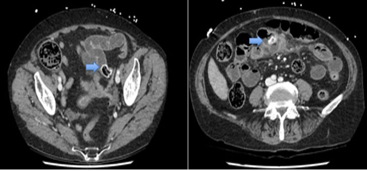
He required emergency dialysis and subsequently was subjected for bilateral nephrostomy to relief the obstruction (Figure 3). During the admission, he was having intermittent fever with no hemodynamic instability observed. After his renal profile and blood gases showed improvement, he was posted for open cystolithotomy (Figure 4).
Intraoperative findings revealed a huge bladder stone measuring 15 cm × 10 cm × 10 cm impacting the whole bladder wall, ureteric opening, and the bladder neck. A moderate amount of pus was present. Initially, the surgeons had difficulty in delivering the bladder stone despite attempting different maneuvers. This is because the stone adhered to the bladder wall. Eventually, the surgeon resorted to fragmentizing the stone into smaller pieces using a chisel and a hammer before it was successfully extracted (Figure 5). During the procedure, care was taken not to traumatize the bladder wall. The weight of the stone recorded is 1 kg.
In post-operation, the patient was managed in the intensive care unit (ICU) for two days before he was transferred to the general ward (Figure 6). Bilateral nephrostomy was removed on day 22 post-surgery. He was discharged well on day 24 post-operation with a bladder catheter after his kidney function showed marked improvement.


Discussion
Bladder stone incidence accounts for only 5% of all urinary calculus. The predisposing factors for bladder stone formation include obstructions and infections such as bladder outlet obstruction, neurogenic bladder, presence of infravesical foreign substance, bladder diverticula, chronic bacteria, and migration from upper urinary tract stone [1]. Middle-aged men are more prone to have bladder stone. In the index case, there is no discernible risk factor for the patient to develop bladder stone. Although spontaneous development of bladder stone is extremely rare, it has been reported before.
A bladder stone weighing more than 100 g is labeled a giant bladder stone. Giant bladder stones are rare today due to better modalities in detecting stones and timely interventions. To cite, fewer than 20 cases of bladder stones were reported to weigh up to 1 kg based on the literature review by Abubakar et al in 2019 [3]. The biggest one found was reported by Goumas et al. (2016), weighing 2.4 kg [4]. In our index case, the weight of the stone is 1 kg.
The association of bladder stones with acute kidney injury is very uncommon. In most of the cases presented with renal dysfunction, it is postulated that the exertion of the relatively large bladder stone on the bladder neck and the mechanical compression on bilateral ureteric orifices will lead to the development of hydronephrosis and eventually, renal impairment [5]. Only a handful of case reports reported the development of kidney failure due to bladder stone as reviewed by Komeya et al. in 2013 [5]. In our case, the chronic obstruction was caused by the large stone that occupied the whole bladder wall. This leads to the stasis and back flow of urine, causing renal impairment. A bilateral nephrostomy tube was placed as a measure to relief the obstruction and the infected system temporarily. This step was taken based on the ultrasound finding before beginning the definitive management of open cystolithotomy.
The treatment of bladder stones depends on the size and the composition of the stone, the comorbidities of the patient, the presence of previous surgery, and anatomic abnormalities of the lower urinary tract, costs, as well as the equipment available. It is essential to eliminate the causative factors in order to ensure therapeutic success [6]. Open cystolithotomy is still the preferred type of surgery for bladder stones measuring more than 4 cm as proposed by Torrecelli and colleague [7]. Though we are moving toward the era of minimally invasive procedures, methods such as extracorporeal shock wave lithotripsy and transurethral cystolithotripsy still has its limitations. The prolonged surgery increases the risk of complications such as urethral stenosis, urethral trauma, and visual disturbance due to hematuria and stone dust [8]. This happens when the stone burden is so large. However, there are several reports of endoscopic and laparoscopic removal of giant bladder calculi. But in these cases, the calculi usually weigh less than 500 g [9],[10].
In our case, open cystolithotomy was performed on the patient. The surgeon encountered difficulty in retrieving the entrapped stone in the bladder. This is due to the size and weight of the stone which impacted the bladder wall, eventually, a chisel was used to break the stone into smaller fragments. Caution was taken to avoid trauma to the bladder mucosa. The stone fragments were extracted with no residual stones left. Chisels are designed to excise bone during orthopedic surgical procedures. Breaking the stone with a chisel and a hammer is a wise alternative in times like these, when the appropriate urological surgery equipment is unavailable. However, it might increase the risk of bladder mucosa injury. By performing the procedure carefully, the surgeon managed to avoid any major complications.
Conclusion
Finding giant bladder stones is an uncommon occurrence. Minimal data exists on the causes, incidence, and management of giant bladder stones, considering its rare occurrence. Further data on the safety of using chisels to break bladder stones should be reviewed.
REFERENCES
1.
Schwartz BF, Stoller ML. The vesical calculus. Urol Clin North Am 2000;27(2):333–46. [CrossRef]
[Pubmed]

2.
Arthure H. A large abdominal calculus. J Obstet Gynaecol Br Emp 1953;60(3):416. [CrossRef]
[Pubmed]
3.
Abubakar BM, Abubakar A, Saleh YM, Suleiman IE. Giant bladder calculus: Case report and literature review. Borno Medical Journal 2019;16(2)
4.
Goumas G, Stasinopoulos K, Fragkoulis C, Papadopoulos G, Stathouros G, Ntoumas K. Giant vesico-prostatic calculus combined with vesicocutaneous fistula. Urol J 2016;13(2):2650–2.
[Pubmed]
5.
Komeya M, Sahoda T, Sugiura S, Sawada T, Kitami K. A huge bladder calculus causing acute renal failure. Urolithiasis 2013;41(1):85–7. [CrossRef]
[Pubmed]
6.
Papatsoris AG, Varkarakis I, Dellis A, Deliveliotis C. Bladder lithiasis: From open surgery to lithotripsy. Urol Res 2006;34(3):163–7. [CrossRef]
[Pubmed]
7.
Torricelli FCM, Mazzucchi E, Danilovic A, Coelho RF, Srougi M. Surgical management of bladder stones: Literature review. Rev Col Bras Cir 2013;40(3):227–33. [CrossRef]
[Pubmed]
8.
Aron M, Agarwal MS, Goel A. Comparison of percutaneous with transurethral cystolithotripsy in patients with large prostates and large vesical calculi undergoing simultaneous transurethral prostatectomy. BJU Int 2003;91(3):293–5. [CrossRef]
[Pubmed]
9.
Nang R, Hinchi H, Lafia T, Rami M, Belkacem R. Giant vesical lithiasis, complication of enterocystoplasty: Case report. Pan Afr Med J 2018;31:132. [CrossRef]
[Pubmed]
10.
Singh I, Hudson JE, Hemal AK. Robot-assisted laparoscopic prostatectomy for a giant prostate with retrieval of vesical stones. Int Urol Nephrol 2010;42(3):615–9 [CrossRef]
[Pubmed]
SUPPORTING INFORMATION
Author Contributions
Mohd Rashid Mazlan - Conception of the work, Design of the work, Acquisition of data, Analysis of data, Drafting the work, Revising the work critically for important intellectual content, Final approval of the version to be published, Agree to be accountable for all aspects of the work in ensuring that questions related to the accuracy or integrity of any part of the work are appropriately investigated and resolved.
Bukhairi Azmi - Conception of the work, Design of the work, Drafting the work, Final approval of the version to be published, Agree to be accountable for all aspects of the work in ensuring that questions related to the accuracy or integrity of any part of the work are appropriately investigated and resolved.
Rashide Yaacob - Revising the work critically for important intellectual content, Final approval of the version to be published, Agree to be accountable for all aspects of the work in ensuring that questions related to the accuracy or integrity of any part of the work are appropriately investigated and resolved.
Guarantor of SubmissionThe corresponding author is the guarantor of submission.
Source of SupportNone
Consent StatementWritten informed consent was obtained from the patient for publication of this article.
Data AvailabilityAll relevant data are within the paper and its Supporting Information files.
Conflict of InterestAuthors declare no conflict of interest.
Copyright© 2022 Mohd Rashid Mazlan et al. This article is distributed under the terms of Creative Commons Attribution License which permits unrestricted use, distribution and reproduction in any medium provided the original author(s) and original publisher are properly credited. Please see the copyright policy on the journal website for more information.


