 |
Case Report
Primary hyperparathyroidism in a giant parathyroid adenoma: A first case report in Jazan, Southern Region, Saudi Arabia
1 Department of Surgery, Jazan University Hospital, Jazan, Saudi Arabia
2 Department of Surgery, King Fahad Central Hospital, Jazan, Saudi Arabia
3 Medical Student, Jazan Medical College, Jazan University, Jazan, Saudi Arabia
Address correspondence to:
Wafa Khudier
Department of Surgery, King Fahad Central Hospital, Jazan,
Saudi Arabia
Message to Corresponding Author
Article ID: 100146Z12HA2024
Access full text article on other devices

Access PDF of article on other devices


How to cite this article
Alessa H, Khudier W, Daak L, Aljudayba R. Primary hyperparathyroidism in a giant parathyroid adenoma: A first case report in Jazan, Southern Region, Saudi Arabia. J Case Rep Images Surg 2024;10(2):32–35.ABSTRACT
Introduction: Primary hyperparathyroidism (PHPT) is the third most prevalent endocrine disorder and is primarily caused by parathyroid adenomas. Giant parathyroid adenoma (GPA), defined as an adenoma weighing over 3.5 g and larger than 2 cm, is a rare entity.
Case Report: This report presents a case of a 70-year-old female from Jazan, Saudi Arabia, diagnosed with GPA and PHPT. The patient exhibited high serum calcium levels and a palpable neck mass, prompting further investigations. Imaging and laboratory findings confirmed the diagnosis, leading to successful surgical removal of the adenoma.
Conclusion: Postoperatively, the patient’s condition improved significantly, highlighting the importance of considering GPA in the differential diagnosis of neck masses.
Keywords: Giant adenoma, Hypercalcemia, Hyperparathyroidism, Parathyroid
Introduction
Primary hyperparathyroidism (PHPT) is a common endocrine disorder characterized by the overproduction of parathyroid hormone (PTH), leading to hypercalcemia. It is most frequently caused by a parathyroid adenoma, which accounts for 80–90% of cases. The prevalence of PHPT is approximately 22 cases per 100,000 individuals, with a higher incidence in postmenopausal women [1]. Although parathyroid carcinoma is a rare cause of PHPT, it should be considered when there is a significantly elevated serum calcium level or a palpable neck mass.
Giant parathyroid adenoma (GPA) is a particularly rare form of parathyroid adenoma, defined by its large size, typically weighing more than 3.5 g and exceeding 2 cm in diameter. The average parathyroid gland weighs between 70 mg and 1 g. GPAs can cause more pronounced clinical symptoms due to their size and the resulting anatomical changes [2],[3].
This report details the first documented case of GPA in the Jazan region of Saudi Arabia. The patient, a 70-year-old female with a history of hypertension, presented with elevated calcium levels and a palpable neck mass. Diagnostic imaging and laboratory tests confirmed the presence of a GPA. Surgical intervention led to the successful removal of the adenoma, and the patient’s postoperative course was uneventful. This case underscores the importance of recognizing GPA as a potential diagnosis in patients with neck masses and elevated calcium levels [4].
Case Report
A 70-year-old female with a known history of hypertension presented to the endocrine surgery clinic with incidental findings of high calcium level during her routine blood workup at her primary health care one year ago, with a history of generalized bone pain and anterior neck swelling.
In the clinic, her physical examination was unremarkable apart from a 3 cm palpable anterior neck mass, on the left side. No other palpable masses.
Laboratory investigations showed a white blood cell count of 5000/µL, hemoglobin (Hb) of 13 g/dL, hematocrit of 40%, and a platelet count of 283×109/L. There was a mildly elevated creatinine level of 119.9 umol/L, blood urea nitrogen (BUN) of 9.2 mmol/L, potassium of 4.1 mmol/L, sodium of 134 mmol/L, and calcium of 3.08 mmol/L, and normal liver function tests and a normal coagulation profile.
Parathyroid hormone level was 151 pmol/L, vitamin D3 was 21.5, phosphorus of was 0.75, thyroid function test showed a thyroid-stimulating hormone (TSH) level of 2.03 mIU/L, T4 of 11.4 mmol/L, and albumin was 39 g/L.
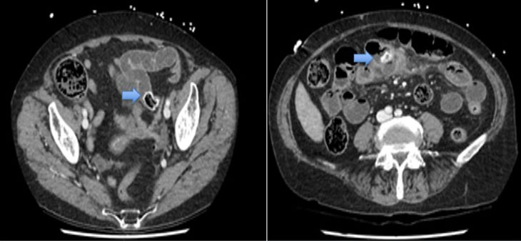
Electrocardiogram (ECG) showed normal sinus rhythm. A neck ultrasound study was done and showed in Figure 1A and Figure 1B.
A large isoechoic left inferior retrothyroid well-defined oval shape mass lesion/nodule measuring about 4.2 × 2.1 × 1.8 cm.
Both thyroid lobes and the thyroid isthmus were average in size with regular outlines, showing homogeneous parenchymal echotexture.
Nuclear study
The thyroid gland showed homogeneous tracer uptake in early images with complete washout in delayed images (Figure 2A).
There was a focus on increased uptake seen posterior to the lower pole of the left thyroid lobe. This was persistent on delayed images.
Scintigraphy evidence of parathyroid adenoma localized inferior to the left thyroid lobe (Figure 2B and Figure 2C).
Dexa scan show
Lumbar spine bone mineral density (BMD) was 0.934 g/cm2, which represented a T-score of –1.5. Left femoral neck BMD was 0.829 g/cm2, which represented a T-score of –0.7.
Right femoral neck BMD was 0.785 g/cm2, which represented a T-score of –1.1. Diagnostic category: Osteopenia according to WHO classification.
The possibility of a parathyroid carcinoma was high enough, based on the patient’s lab and imaging findings, to recommend the patient’s surgery for tumor removal. The operation was performed uneventfully as a left inferior parathyroidectomy. An intraoperative level of PTH was obtained and showed a 50% drop off compared to the preoperative level. The postoperative course was uneventful. The patient was discharged home on day 2.
Histopathology result
Left inferior parathyroid gland/Excision: Specimen photograph of the giant parathyroid adenoma.
- A well-circumscribed parathyroid lesion with histologic features in favor of parathyroid adenoma.
- The sections of the parathyroid gland show an adenoma consisting predominantly of chief cells and some clear cells.
Normal parathyroid tissue admixed with adipose tissue is seen adjacent to the adenoma.
- No necrosis or atypical mitosis.


Discussion
Primary hyperparathyroidism (PHP) is the third most common endocrine disorder and is usually due to parathyroid adenoma. The disease usually presents as mild asymptomatic hypercalcemia discovered during routine biochemical testing.
Parathyroid carcinoma is the least common cause of PHP but should be considered in the differential diagnosis of PHP when the serum calcium (mg/dL) level is above 14 or PHP is associated with a palpable neck mass.
However, giant parathyroid adenoma can cause symptoms due to pressure effect, including dysphagia, odynophagia, and dyspnea. Clinical studies show that hypercalcemia crisis is very common emergency presenting with palpable parathyroid mass. Biochemical increase in calcium levels and parathyroid (PTH) function facilitate the diagnosis. Radiological ultrasonography and 99mTc-MIBI scintigraphy facilitate the localization and characterization of giant parathyroid adenoma.
This patient had been suffering from a palpable neck mass with mild symptoms of hypercalcemia. Considering her back and bone pain, the adenoma might have been chronically functional. Diagnosis of a parathyroid mass was made based on lab findings, including high calcium and PTH levels. The level of PTH was almost 70 times its expected value.
With this high level of PTH and the presence of palpable neck mass, parathyroid carcinoma was suspected, and surgical excision was indicated.
Intraoperatively, there are no suspicious lymph nodes or other neck mass apart from the left inferior large parathyroid adenoma, which was resected, and specimen sent for histopathology. Intraoperative PTH level dropped more than 50% after taking of adenoma. The postoperative course was uneventful, the laboratory workup was unremarkable, and the patient was discharged home on day 2 with oral calcium and vitamin D3 supplements.
The patient followed in outpatient department (OPD) and had an uneventful postoperative course. The patient has no symptoms of hypo- or hyperparathyroidism and normal calcium level with oral supplements for several months.
In summary, hyperparathyroidism should be considered in the differential diagnosis of any neck and cervical mass, even though it is a sporadic tumor and parathyroid carcinoma should be excluded in case of a giant parathyroid tumor.
Conclusion
The definition or cut-off for a giant parathyroid adenoma is still an open issue; however, the values used so far include a weight of >3–3.5 g and size of more than 2 cm. Giant adenomas are primarily associated with primary hyperparathyroidism (PHPT), exhibiting higher levels of calcium and PTH than non-giant adenomas, but typically resulting in fewer complications. This may be due to the presence of a neck mass, which prompts patients to seek medical attention earlier. In these conditions, surgical excision is necessary to treat the symptoms of hyperparathyroidism and rule out other possibilities of parathyroid carcinoma.
REFERENCES
1.
Mahmodlou R, Sedokani A, Pezeshk A, Najafinejad B. Giant parathyroid adenoma: A case report. J Med Case Rep 2022;16(1):150. [CrossRef]
[Pubmed]

2.
Krishnamurthy A, Raghunandan GC, Ramshankar V. A rare case of giant parathyroid adenoma presenting with recurrent episodes of pancreatitis. Indian J Nucl Med 2016;31(1):36–8. [CrossRef]
[Pubmed]
3.
Ghemigian A, Trandafir AI, Petrova E, et al. Primary hyperparathyroidism-related giant parathyroid adenoma (Review). Exp Ther Med 2022;23(1):88. [CrossRef]
[Pubmed]
4.
Bhan A, Athimulam S, Kumari P, et al. Large parathyroid adenomas: Potential mechanisms to reconcile adenoma size and disease phenotype. Front Endocrinol (Lausanne) 2023;14:1009516. [CrossRef]
[Pubmed]
SUPPORTING INFORMATION
Author Contributions
Halimah Alessa - Conception of the work, Design of the work, Acquisition of data, Analysis of data, Drafting the work, Revising the work critically for important intellectual content, Final approval of the version to be published, Agree to be accountable for all aspects of the work in ensuring that questions related to the accuracy or integrity of any part of the work are appropriately investigated and resolved.
Wafa Khudier - Conception of the work, Design of the work, Acquisition of data, Analysis of data, Drafting the work, Revising the work critically for important intellectual content, Final approval of the version to be published, Agree to be accountable for all aspects of the work in ensuring that questions related to the accuracy or integrity of any part of the work are appropriately investigated and resolved.
Lojain Daak - Analysis of data, Drafting the work, Final approval of the version to be published, Agree to be accountable for all aspects of the work in ensuring that questions related to the accuracy or integrity of any part of the work are appropriately investigated and resolved.
Reham Aljudayba - Analysis of data, Drafting the work, Final approval of the version to be published, Agree to be accountable for all aspects of the work in ensuring that questions related to the accuracy or integrity of any part of the work are appropriately investigated and resolved.
Guarantor of SubmissionThe corresponding author is the guarantor of submission.
Source of SupportNone
Consent StatementWritten informed consent was obtained from the patient for publication of this article.
Data AvailabilityAll relevant data are within the paper and its Supporting Information files.
Conflict of InterestAuthors declare no conflict of interest.
Copyright© 2024 Halimah Alessa et al. This article is distributed under the terms of Creative Commons Attribution License which permits unrestricted use, distribution and reproduction in any medium provided the original author(s) and original publisher are properly credited. Please see the copyright policy on the journal website for more information.

