|
Case Report
Strangulated Richter’s femoral hernia in a male patient: A rare and atypical presentation
1 Department of General Surgery, Arrowhead Regional Medical Center, Colton, CA 92324, United States of America
2 School of Medicine, St-George’s University, True Blue, St. George’s, Grenada, West Indies
Address correspondence to:
Elias Wassel
MD, Arrowhead Regional Medical Center, 400 N Pepper Ave, Colton, CA 92324,
United States America
Message to Corresponding Author
Article ID: 100172Z12EW2026
Access full text article on other devices

Access PDF of article on other devices


How to cite this article
Wassel E, Hormozian S, Lee M, Sanchez L, Robinson N. Strangulated Richter’s femoral hernia in a male patient: A rare and atypical presentation. J Case Rep Images Surg 2026;12(2):10–13.ABSTRACT
Introduction: Richter’s hernia is an uncommon hernia in which the anti-mesenteric bowel wall becomes entrapped within a small fascial defect. Because intestinal continuity is preserved, patients may pass stool despite ongoing ischemia, often delaying diagnosis. Richter’s hernias most commonly occur in narrow anatomical openings, and are more common in females, making this presentation in a male patient particularly rare.
Case Report: A 78-year-old male with dementia presented with four days of abdominal pain and poor oral intake. Imaging demonstrated small bowel obstruction due to a right groin hernia with surrounding edema. Urgent surgery identified a strangulated Richter’s femoral hernia with a 3-cm hemorrhagic, edematous anti-mesenteric bowel segment requiring resection and anastomosis. McVay tissue repair was performed. The patient recovered well and was discharged on postoperative day five.
Conclusion: This case highlights the importance of maintaining a high index of suspicion for Richter’s femoral hernia in patients with unexplained groin pain to prevent ischemia, necrosis, and associated morbidity.
Keywords: Femoral hernia, Richter’s hernia, Small bowel obstruction, Strangulated hernia
Introduction
Richter’s hernia is an uncommon but clinically significant type of hernia in which only the anti-mesenteric portion of the bowel wall becomes entrapped within a small, rigid fascial defect. Because only part of the bowel circumference is involved, intestinal continuity is preserved, allowing patients to continue passing flatus and stool despite ongoing vascular compromise. This lack of complete obstruction frequently leads to delayed diagnosis, with many cases presenting only after ischemia or gangrene of the involved bowel has developed.
Richter’s hernias most commonly occur at narrow anatomical openings with firm margins, particularly the femoral canal. Femoral hernias account for approximately 2–4% of all groin hernias and carry a high risk of incarceration and strangulation [1]. Although femoral hernias are more common in females, Richter’s femoral hernia should still be considered in male patients presenting with compatible symptoms, despite its rarity.
The first documented case of Richter’s hernia was reported by Fabricius Hildanus in 1558, but it was not until 1778 that German surgeon August Gottlieb Richter provided the first scientific description of the condition, which ultimately came to bear his name [2]. The presence of a tight constricting ring is a prerequisite for strangulation and compromised blood circulation, ultimately leading to ischemia and gangrene of the involved bowel [3]. Richter’s hernia may occur at any typical hernia site but is most likely to develop in small hernial rings with firm margins.
Reported series suggest that 36–88% of Richter’s hernias occur within the femoral canal, followed by the inguinal canal and incisional hernias [2]. Richter’s femoral hernias are rare and can present without classic obstructive symptoms, creating significant diagnostic challenges. This case highlights the diagnostic challenges associated with atypical presentations and underscores the importance of maintaining clinical suspicion despite the absence of classic obstructive symptoms [4].
Case Report
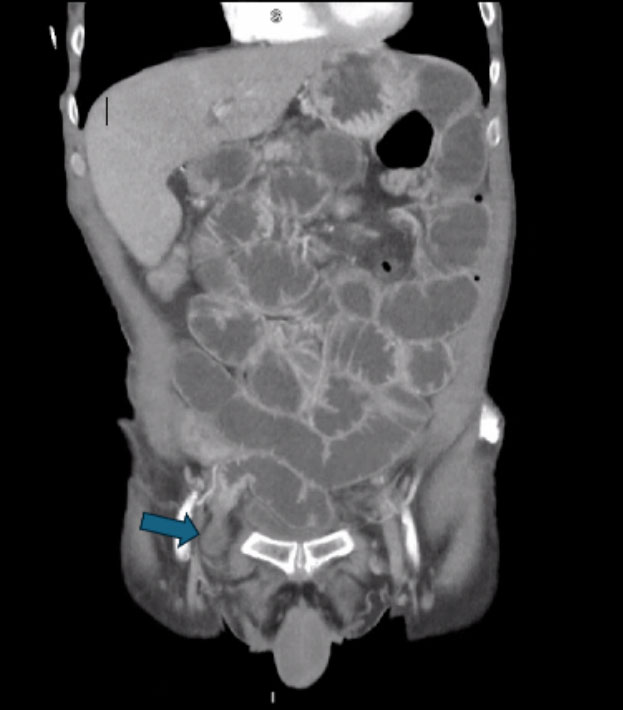
A 78-year-old male with a past medical history of dementia, hypertension, hyperlipidemia, hypothyroidism, chronic obstructive pulmonary disease, and a recent stroke presented with right-sided abdominal pain of four days’ duration. During this time, he was partially able to tolerate oral intake and experienced multiple episodes of nausea and vomiting. He also reported intermittent bowel movements over the two days preceding presentation. Computed tomography (CT) abdomen and pelvis demonstrated a small bowel obstruction likely secondary to a right groin hernia containing a focal loop of small bowel, with a clear transition point consistent with an incarcerated groin hernia (Figure 1). The herniated segment protrudes through a narrow femoral defect and appears to involve only part of the bowel wall rather than the full circumference; findings were suspicious for a Richter-type hernia. Surrounding edema and mild bowel wall thickening raise concern for early ischemic changes. Laboratory evaluation was significant for leukocytosis (16.5 × 10³ µL), elevated lactate (3.44 mmol/L), and acute kidney injury (creatinine 2.55 mg/dL). On examination, the patient demonstrated localized right groin tenderness with a palpable, firm, small, irreducible hernia mass, without overlying skin changes. The abdomen was soft and nondistended. Vital signs were unremarkable.
The patient was taken urgently to the operating room, where a femoral hernia was identified with strangulation of only the anti-mesenteric portion of the small bowel through the canal defect, consistent with a Richter’s hernia. The affected 3 cm segment appeared dark red, hemorrhagic, and edematous. Resection of the involved segment was performed, followed by a side-to-side jejuno-jejunal anastomosis (jejunojejunostomy). The femoral hernia defect was repaired using a McVay tissue repair.
Postoperatively, the patient’s recovery was uneventful. The patient had full return of bowel function on post-operative day four and was discharged home. Histopathology of the resected segment demonstrated focal transmural hemorrhage with ischemic changes. The patient has since been seen in clinic and continues to recover as expected.
Discussion
Clinically, Richter hernias can be difficult to diagnose, as patients may not present with complete bowel obstruction, similar to our patient. Subtle symptoms, such as localized pain or intermittent vomiting, can delay recognition, increasing the risk of ischemia. Imaging, particularly CT, is valuable in identifying the partially entrapped bowel loop and guiding timely surgical intervention [5].
In our case, the patient presented with right-sided abdominal pain and intermittent episodes of vomiting, with some oral intake, prompting surgical intervention based on the CT findings. Operative management involved releasing the incarcerated bowel loop and resecting the necrotic segment, which aligns with literature reporting a high incidence of bowel necrosis—up to 69%—at the time of intervention in Richter’s hernias [2]. The anti-mesenteric portion of the bowel is particularly prone to necrosis due to its limited collateral blood supply, explaining the progression to ischemia observed in this patient, a finding corroborated by histopathology showing transmural hemorrhage of the bowel wall (Figure 2).
This case suggests that favorable outcomes may still occur despite delayed presentation when early imaging diagnosis, expedited surgery, and limited bowel involvement allow intervention prior to transmural necrosis or perforation.
Given the characteristics of the hernia and the patient’s presentation, we performed an open McVay (Cooper’s ligament) repair to provide optimal exposure and facilitate secure closure of the femoral defect. Although minimally invasive approaches, including robotic and laparoscopic repair, are feasible, the open technique was selected in anticipation of partial division of the inguinal ligament to ensure adequate exposure and safe reduction of the herniated bowel. Postoperative outcomes are generally favorable when prompt intervention is performed.

Conclusion
This case highlights the diagnostic challenge of Richter’s femoral hernia due to the absence of complete bowel obstruction and nonspecific early findings. Prompt recognition, a high index of suspicion, and timely surgical intervention are essential to prevent ischemia, necrosis, and associated complications. Clinicians should consider this diagnosis in patients presenting with unexplained abdominal or groin pain, regardless of sex.
REFERENCES
1.
Fitzgibbons RJ Jr, Forse RA. Clinical practice. Groin hernias in adults. N Engl J Med 2015;372(8):756–63. [CrossRef]
[Pubmed]

2.
Steinke W, Zellweger R. Richter’s hernia and Sir Frederick Treves: An original clinical experience, review, and historical overview. Ann Surg 2000;232(5):710–8. [CrossRef]
[Pubmed]
3.
Horbach JM. Invagination for Richter-type strangulated hernias. Trop Doct 1986;16(4):163–8. [CrossRef]
[Pubmed]
4.
Parmar VS, Vaghasiya GN, Dave JP. A rare case of Richter’s variety of obstructed femoral hernia in a male. Indian J Surg 2015;77(Suppl 1):79–80. [CrossRef]
[Pubmed]
5.
Smith CR, Chatzikonstantinou M. Early surgical intervention is critical for strangulated Richter’s hernia. J Surg Case Rep 2024;2024(10):rjae642. [CrossRef]
[Pubmed]
SUPPORTING INFORMATION
Author Contributions
Elias Wassel - Conception of the work, Design of the work, Drafting the work, Revising the work critically for important intellectual content, Final approval of the version to be published, Agree to be accountable for all aspects of the work in ensuring that questions related to the accuracy or integrity of any part of the work are appropriately investigated and resolved.
Sean Hormozian - Conception of the work, Design of the work, Acquisition of data, Analysis of data, Drafting the work, Final approval of the version to be published, Agree to be accountable for all aspects of the work in ensuring that questions related to the accuracy or integrity of any part of the work are appropriately investigated and resolved.
Michelle Lee - Conception of the work, Design of the work, Drafting the work, Revising the work critically for important intellectual content, Final approval of the version to be published, Agree to be accountable for all aspects of the work in ensuring that questions related to the accuracy or integrity of any part of the work are appropriately investigated and resolved.
Luciano Sanchez - Conception of the work, Design of the work, Acquisition of data, Analysis of data, Revising the work critically for important intellectual content, Final approval of the version to be published, Agree to be accountable for all aspects of the work in ensuring that questions related to the accuracy or integrity of any part of the work are appropriately investigated and resolved.
Nia Robinson - Conception of the work, Design of the work, Drafting the work, Revising the work critically for important intellectual content, Final approval of the version to be published, Agree to be accountable for all aspects of the work in ensuring that questions related to the accuracy or integrity of any part of the work are appropriately investigated and resolved.
AcknowledgmentsThe authors would like to acknowledge the contributions of the radiology, and pathology team for their role in the diagnosis and management of this case. They also thank the patient and his family for their consent to publish this case report (IRB #26-11).
Artificial intelligence (AI) use in the article
I certify that AI tools were used only for drafting or language assistance in preparation of the manuscript, and that all facts, data, and records presented in the manuscript are the authors’ own intellectual contribution, and no part of the facts, data, and records presented in the manuscript, are generated using AI tools.
The corresponding author is the guarantor of submission.
Source of SupportNone
Consent StatementWritten informed consent was obtained from the patient for publication of this article.
Data AvailabilityAll relevant data are within the paper and its Supporting Information files.
Conflict of InterestAuthors declare no conflict of interest.
Copyright© 2026 Elias Wassel et al. This article is distributed under the terms of Creative Commons Attribution License which permits unrestricted use, distribution and reproduction in any medium provided the original author(s) and original publisher are properly credited. Please see the copyright policy on the journal website for more information.